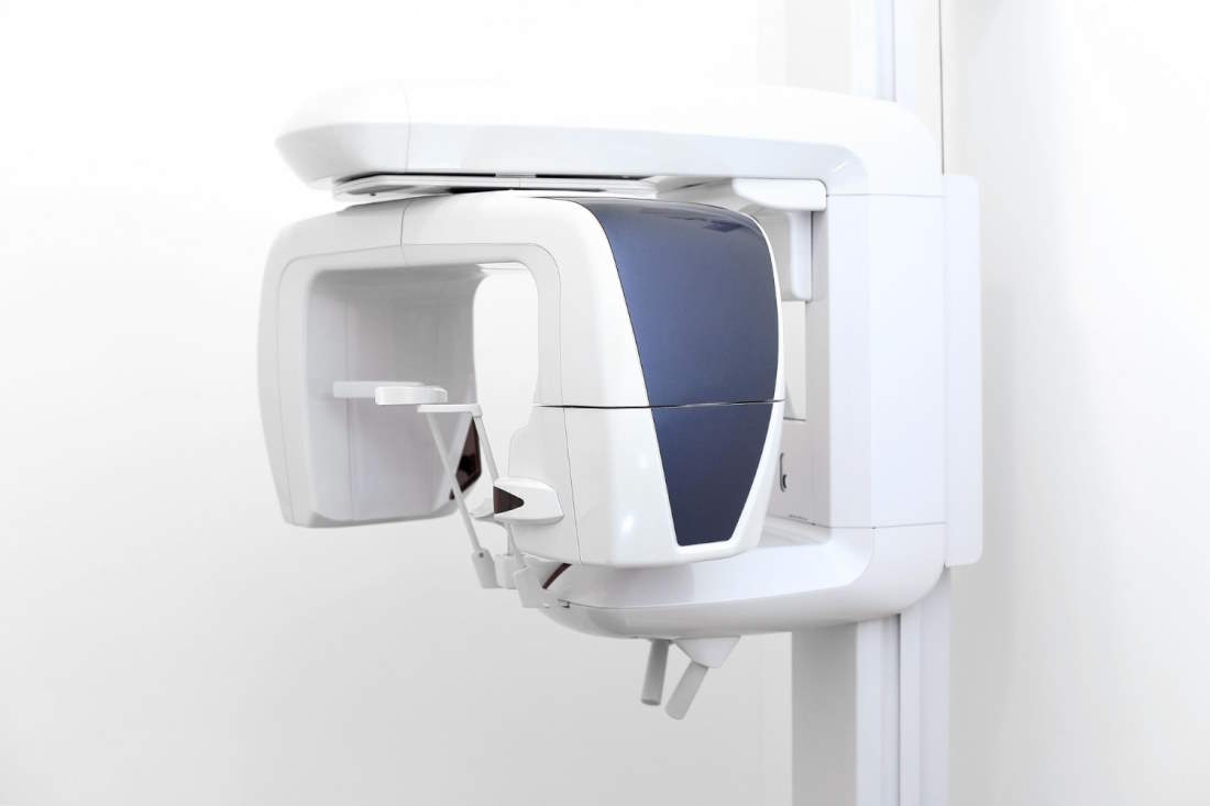
Digital Panoramic Radiography is the most useful examination in dental practice. It is a radiography that displays all the teeth and the bones of the jaws, the sinuses, the nasal cavity, and the cervical tissues with exceptional clarity. It is used for the diagnosis of dental problems, allowing the doctor to have a full view of the teeth in one image in order to assess any issues and determine the appropriate treatment. At the same time, it displays previous dental issues that were addressed, such as root canals and fillings.
Cephalometric Digital Radiography
Cephalometric Digital Radiography is the digital radiography that displays the head in a special lateral position to assess the distances, structure, and position of the bones and soft tissues of the skull.
The examination is conducted in a standing position, with examinees biting down on a plastic piece for a few seconds while the machine (arm – lamp) makes a complete rotation around their head. The duration of the examination is a few seconds, and the examinee must remain still in the position indicated by the technologist.
CBCT (Cone Beam Computed Tomography)
Cone Beam Computed Tomography (CBCT) is considered one of the most significant inventions in the field of imaging and diagnosis in modern dentistry. It is a new radiographic technique where the machine rotates around the patient's head, producing over six hundred (600) images, which are then reconstructed into a three-dimensional (3D) image using specialized software. This method achieves exceptionally high-quality and precise three-dimensional imaging of the structures of the jaws and teeth. Additionally, the patient is exposed to a much lower dose of radiation compared to conventional computed tomography. Consequently, Cone Beam Computed Tomography has become the most modern and significant diagnostic method in dentistry, gaining ground in clinical practice every day.
Areas in which three-dimensional Cone Beam Computed Tomography (CBCT) is used:
- Implants: In preoperative planning for implant placement: (a) identification and diagnosis of significant anatomical landmarks, such as the inferior alveolar nerve, (b) three-dimensional imaging of the shape and morphology of the ridge, (c) assessment of bone quality, and (d) determination of implant placement positions.
- Impacted canines, wisdom teeth
- Cysts, abscesses, neoplasms
- Apical lesions not identified in Panoramic radiographs.
- Endodontics
- Orthodontics
- Periodontology


