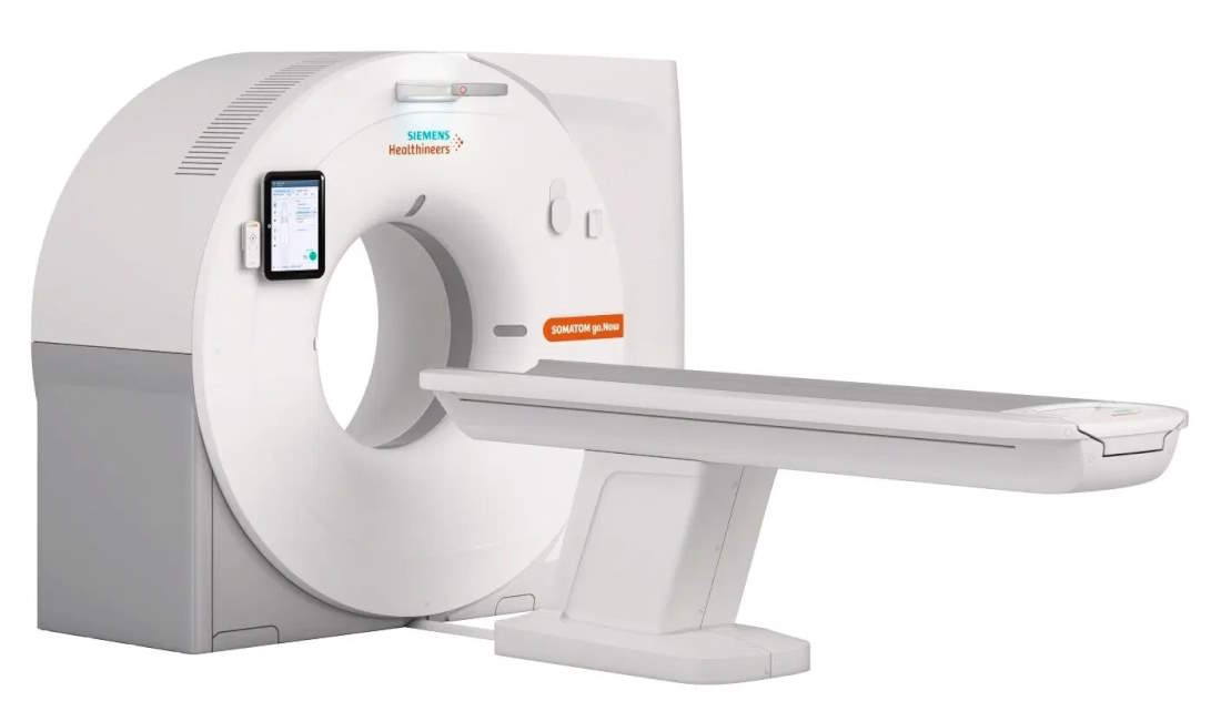
Computed Tomography (CT) is a highly analytical imaging procedure that is performed using X-rays and can accurately depict many parts of the body, including the brain, lungs, bones, soft tissues, the heart, and even the oral cavity.
The CT scan examination is painless and of short duration, as with modern CT scanners it can be completed within a timeframe ranging from 5 to 20 minutes, including the required preparation time. During the examination, and depending on its type, a contrast agent may be administered for better imaging of the area.
CT Angiography
CT angiography is performed on a multislice CT scanner after the intravenous injection of a contrast agent. The contrast agent is later excreted by the kidneys into the urine. This is a painless examination that lasts only a few minutes. The examination checks the vessels of the brain, neck, upper and lower limbs, as well as all body regions (thoracic aorta, abdominal aorta, renal arteries, iliac arteries, etc.). CT angiography is a modern application in the investigation of the body and head vessels. With this examination, we can image the vessels in any area of the human body in a non-invasive, painless, bloodless way, avoiding the relatively painful and not complication-free traditional angiography. This way, we can diagnose stenoses, obstructions, aneurysms, or other vascular diseases with great diagnostic accuracy.
CT angiography is recommended in the following cases:
- Diseases of the brain vessels (aneurysms, malformations, stenoses, obstructions) and the carotids
- Aortic diseases (aneurysms, stenoses, dissecting aneurysms, post-stent placement check)
- Examination of the upper limbs (embolism, trauma) and lower limbs (arterial stenosis or obstruction, venous emboli)
- Examination of the coronary arteries of the heart (stenoses, calcium score estimation)
- Examination of the pulmonary arteries (pulmonary embolism)
- Examination of the liver vessels (trauma, hepatic vein obstruction)
- Examination of the renal vessels
Preparation and conduct of the examination
The examination is quick, easy, and painless. Before the examination, the examinee will be asked to fast for several hours.
The examinee lies on the examination table of the CT scanner. Then, a small catheter is placed in a vein of the hand, which is connected to the contrast injector. Due to the contrast, you may feel warmth from head to toe, which will quickly subside. During the examination, the technologist may ask you to hold your breath for a few seconds. You may also be asked to remain completely still to ensure clear images. The entire examination process usually lasts less than 30 minutes, and the examinee can immediately return to their daily activities.
CT Pyelography
CT pyelography, or urography, is the computed tomography that examines the urinary system, providing useful information not only about the function but also about the morphology of the system. CT pyelography is performed on a multislice CT scanner after the prior intravenous injection of a contrast agent. The contrast agent is later excreted by the kidneys into the urine. It is a painless examination that lasts only a few minutes. With CT pyelography, the kidneys, ureters, urinary bladder, and generally the entire system are examined.
CT pyelography is recommended in the following cases:
- Checking the function of the kidneys
- Checking for cancer of the urinary system (painless hematuria)
- Diagnosis of stones
- Injury to the urinary system, e.g., kidney rupture
- Imaging of distant metastases
- Staging of tumors


